
Katsamenis, Orestis, Warner, Jane, Sinclair, Ian, Lackie, Peter and Schneider, Philipp (2019) Dataset for X-ray micro-computed tomography for non-destructive 3D X-ray histology. University of Southampton doi:10.5258/SOTON/D0902 [Dataset]
Abstract
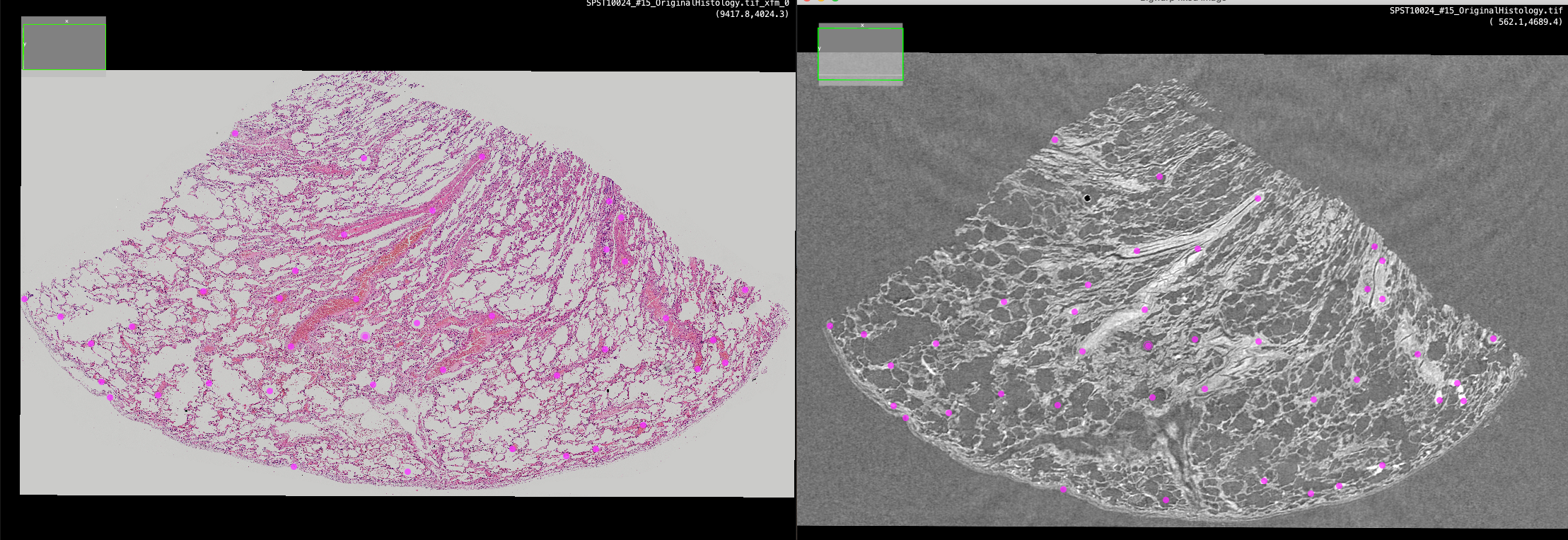
Datasets used for the study entitled 'X-ray micro-computed tomography for non-destructive 3D X-ray histology' by Orestis L. Katsamenis, Michael Olding, Jane A. Warner, David S. Chatelet, Mark G. Jones, Giacomo Sgalla, Bennie Smit, Oliver J. Larkin, Ian Haig, Luca Richeldi, Ian Sinclair, Peter M. Lackie, Philipp Schneider. The American Journal of Pathology Abstract for the paper Historically, micro-computed tomography has been considered unsuitable for histological analysis of unstained formalin-fixed and paraffin-embedded (FFPE) soft tissue biopsies due to a lack of image contrast between the tissue and the paraffin. However, we recently demonstrated that μCT can successfully resolve microstructural detail in routinely prepared tissue specimens. Here, we illustrate how μCT imaging of standard FFPE biopsies can be seamlessly integrated into conventional histology workflows, enabling non-destructive three-dimensional (3D) X-ray histology, the use and benefits of which we showcase for the exemplar of human lung biopsy specimens. This technology advancement was achieved through manufacturing a first-of-kind μCT scanner for X-ray histology and developing optimised imaging protocols, which do not require any additional sample preparation. 3D X-ray histology allows for non-destructive 3D imaging of tissue microstructure, resolving structural connectivity and heterogeneity of complex tissue networks, such as the vascular or the respiratory tract. We also demonstrate that 3D X-ray histology can yield consistent and reproducible image quality, enabling quantitative assessment of tissue’s 3D microstructures, which is inaccessible to conventional two-dimensional histology. Being non-destructive the technique does not interfere with histology workflows, permitting subsequent tissue characterisation by means of conventional light microscopy-based histology, immunohistochemistry, and immunofluorescence. 3D X-ray histology can be readily applied to a plethora of archival materials, yielding unprecedented opportunities in diagnosis and research of disease.
More information
Identifiers

Catalogue record
Export record
Altmetrics
Contributors
Download statistics
Downloads from ePrints over the past year. Other digital versions may also be available to download e.g. from the publisher's website.