Dataset in support of the article 'Laser induced forward transfer imaging using deep learning'
Dataset in support of the article 'Laser induced forward transfer imaging using deep learning'

This dataset contains:
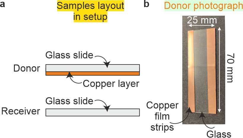
Fig. 1 a) Side view of receiver and donor samples. b) Photograph of donor copper film with blank region where tape was used to hold sample in deposition chamber.
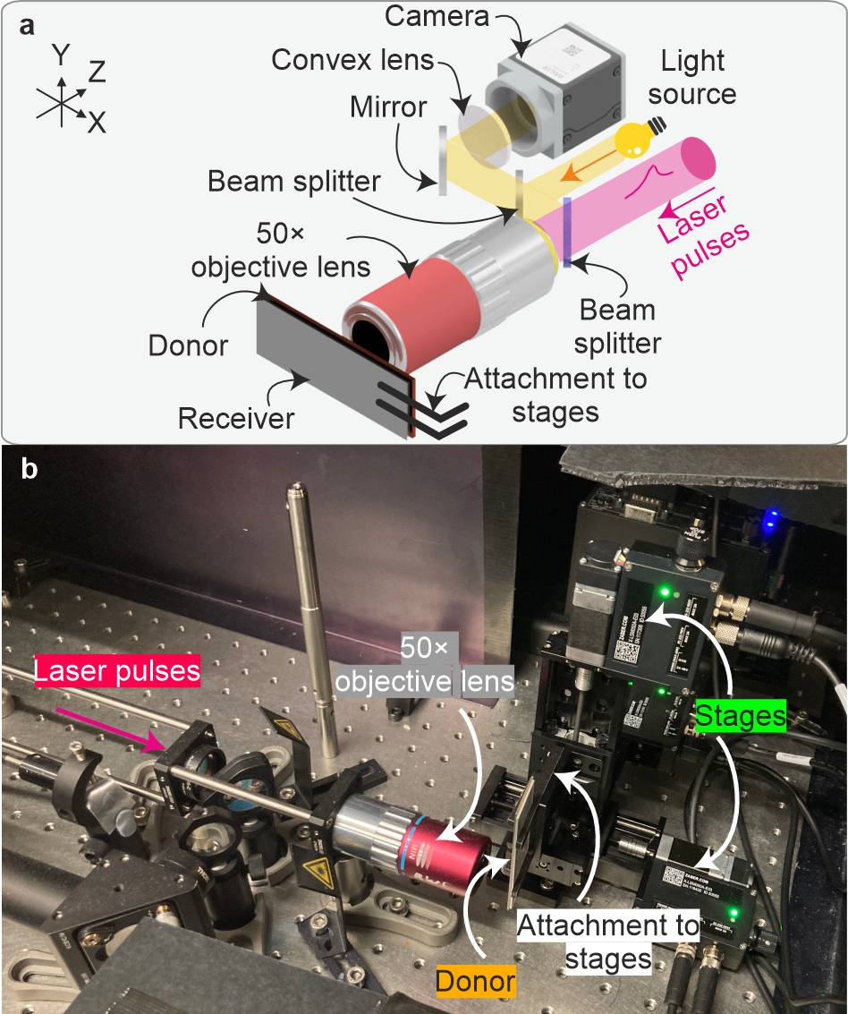
Fig. 2 a) Laser pulses were focussed onto the surface of the donor substrate and copper was deposited onto a receiver. Following depositions, the samples were imaged using a separate Nikon microscope. b) Photograph of the setup with labels indicating key apparatus.
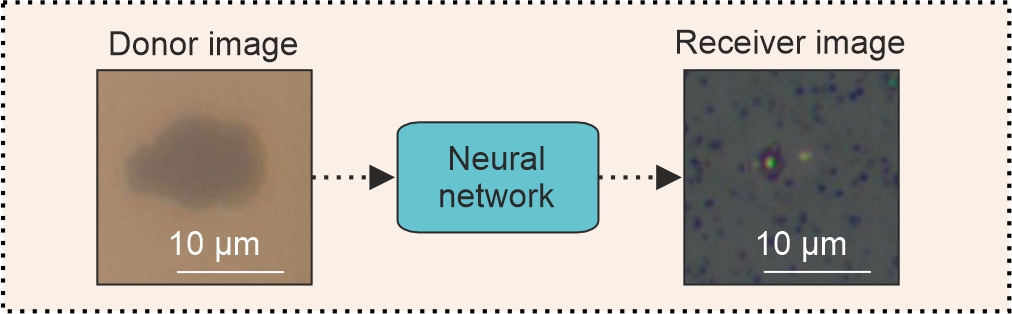
Fig. 3 Schematic concept of feeding the donor image into the neural network to produce an image of the receiver.
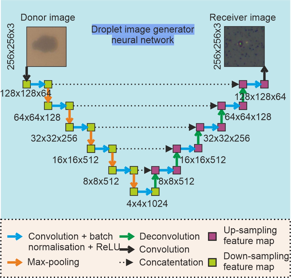
Fig 4. Diagram of the neural network for image generation.
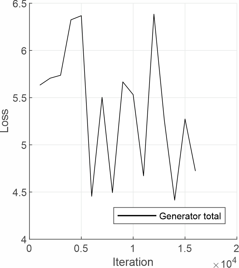
Fig. 5 Total generator loss as a function of neural network training iteration.
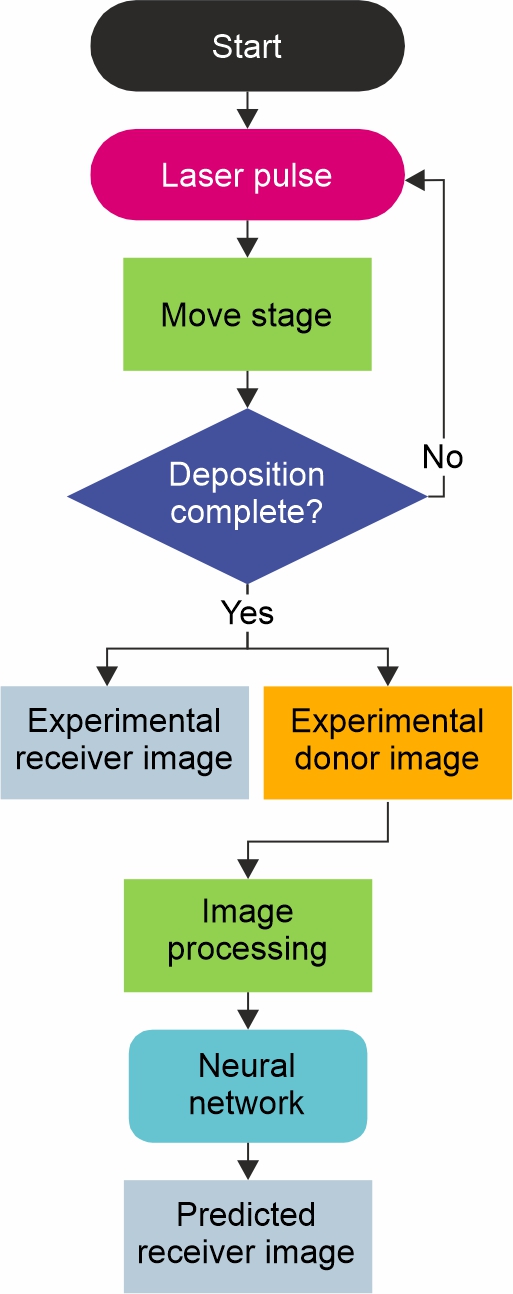
Fig. 6 Flowchart of deposition and image prediction process.
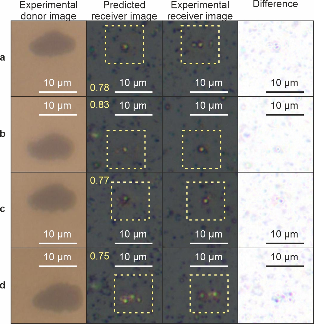
Fig. 7 Predicting the appearance of the receiver from images of the donor for laser pulse fluence of a 5.4 Jcm-2, b 6.1 Jcm-2, c 6.5 Jcm-2 and d 7.1 Jcm-2, with size scales shown in white and SSIM values inset in yellow
Table 1 Laser specifications
Table 2 Microscope Basler camera specifications
Table 3 SSIM and RMSE values for the images shown in Fig. 7.
Article to be published in Discover Applied Sciences
University of Southampton
Grant-Jacob, James A.
c5d144d8-3c43-4195-8e80-edd96bfda91b
Zervas, Michalis
1840a474-dd50-4a55-ab74-6f086aa3f701
Mills, Ben
05f1886e-96ef-420f-b856-4115f4ab36d0
Grant-Jacob, James A.
c5d144d8-3c43-4195-8e80-edd96bfda91b
Zervas, Michalis
1840a474-dd50-4a55-ab74-6f086aa3f701
Mills, Ben
05f1886e-96ef-420f-b856-4115f4ab36d0
Grant-Jacob, James A., Zervas, Michalis and Mills, Ben
(2025)
Dataset in support of the article 'Laser induced forward transfer imaging using deep learning'.
University of Southampton
doi:10.5258/SOTON/D3416
[Dataset]
Abstract
This dataset contains:
Fig. 1 a) Side view of receiver and donor samples. b) Photograph of donor copper film with blank region where tape was used to hold sample in deposition chamber.
Fig. 2 a) Laser pulses were focussed onto the surface of the donor substrate and copper was deposited onto a receiver. Following depositions, the samples were imaged using a separate Nikon microscope. b) Photograph of the setup with labels indicating key apparatus.
Fig. 3 Schematic concept of feeding the donor image into the neural network to produce an image of the receiver.
Fig 4. Diagram of the neural network for image generation.
Fig. 5 Total generator loss as a function of neural network training iteration.
Fig. 6 Flowchart of deposition and image prediction process.
Fig. 7 Predicting the appearance of the receiver from images of the donor for laser pulse fluence of a 5.4 Jcm-2, b 6.1 Jcm-2, c 6.5 Jcm-2 and d 7.1 Jcm-2, with size scales shown in white and SSIM values inset in yellow
Table 1 Laser specifications
Table 2 Microscope Basler camera specifications
Table 3 SSIM and RMSE values for the images shown in Fig. 7.
Article to be published in Discover Applied Sciences
More information
Published date: 2025
Identifiers
Local EPrints ID: 499199
URI: http://eprints.soton.ac.uk/id/eprint/499199
PURE UUID: faeb07b8-45a2-4311-b96f-8ffbde145c9a
Catalogue record
Date deposited: 11 Mar 2025 18:02
Last modified: 12 Mar 2025 02:44
Export record
Altmetrics
Contributors
Creator:
James A. Grant-Jacob

Creator:
Michalis Zervas
Creator:
Ben Mills
Download statistics
Downloads from ePrints over the past year. Other digital versions may also be available to download e.g. from the publisher's website.
View more statistics